Measuring humidity in dynamic situations calls for highly sensitive fast response sensors. Here we report, a humidity sensor fabricated using solution processed supramolecular nanofibres as active resistive sensing material. The nanofibres are built via self- assembly of donor and acceptor molecules (coronene tetracarboxylate and dodecyl methyl viologen respectively) involved in charge transfer interactions. The conductivity of the nanofibre varied sensitively over a wide range of relative humidity (RH) with unprecedented fast response and recovery times. Based on UV-vis, XRD and AFM measurements, it is found that the stacking distance in the nanofibre decreases slightly while the charge transfer band intensity increases, all observations implying enhanced charge transfer interaction and hence the conductivity. It is demonstrated to be as a novel breath sensor which can monitor the respiration rate. Using two humidity sensors, a breath flow sensor was made which could simultaneously measure RH and flow rate of exhaled nasal breath. The integrated device was used for monitoring RH in the exhaled breath from volunteers undergoing exercise and alcohol induced dehydration.
Humidity sensing is important in different settings, in chemical industry, hospitals, agricultural fields and so on 1,2 . A sensor working with fast response and recovery (typically fraction of a second) finds niche applications in situations where the humidity value may change dynamically. Such application areas include industrial process controls, meteorology and various medical diagnostics including monitoring human exhaled breath 3,4,5,6,7 . This explains the need of low cost humidity sensors with ultra-fast response, high sensitivity and stability. Although the literature is abound with humidity sensors 8 , those with fast response form only a small subset (see Supplementary Table S1). The subset includes those based on inorganic materials in the form of nanowires 9 , nanotubes 10,11 , thin films of oxides 12,13,14 and of few sulfides 15,16 as well. Similarly, there are examples from organic and composite materials, a notable one being nafion persulphonate 4 with tens of milliseconds of response time. Very recently, Borini et al. 17 have reported response time of 30 ms for a graphene oxide based sensor. In addition to this, there are advancements in fabrication of humidity sensors using optic fibre based sensor technology 18,19 . There have been reports of humidity sensors using hybrid microcavity 20 , AFM micro-cantilevers 21 , plasmonic materials 22 etc. Fabrication of flexible, printable, chipless and wireless humidity sensors and integrating them with CMOS technology has been recently reported intended for various humidity related applications 23,24,25 . The basic mechanism of humidity detection in the above materials is based on the change in proton/ionic conduction (resistive), dielectric constant (capacitive), refractive index (optical), frequency (impedance) or in mass, of the active material with the humidity level 26 . Organic humidity sensors are particularly attractive since the sensing material can be prepared by simple solution processes unlike in inorganic sensors which demand expensive preparation techniques such as chemical vapor deposition, vapour-liquid-solid growth etc.; however, organic sensors usually lack stability in terms of device performance 27,28,29 .
Supramolecular systems are relatively a new class of organic materials, built via non-covalent interactions among the molecular ingredients 30 . Fabricating functional devices such as FETs, solar cells, sensors from controlled organization of supramolecular components is considered as a promising area of research 31,32,33,34 . It takes the advantage of intermediate size regime, few tens of nm to μm, when compared to single molecule (few nm) and polymer films (≈μm) 35,36 . Recently, Herzer et al. 37 reported change in color of supramolecular polymer films upon exposure to humidity. Among various supramolecular systems, 1-D nanowires are of particularly interesting since they provide low scattering conducting paths 38,39,40 and easy alignment across the electrodes 41 . We have recently embarked on a supramolecular one-dimensional (1-D) system, a charge transfer donor and acceptor couple grown in the form of nanofibre, to use as an active channel material in field-effect transistor which worked in the ambient conditions and showed high hole mobility and carrier concentration 42 . During the measurements, it was observed that the device performance was sensitively dependent on the laboratory humidity conditions. In the present article, we have examined this aspect in detail and developed a humidity sensor based on resistive response of the supramolecular nanofibre at different levels of humidity. The associated structural changes and interactions therein, have been examined using UV-vis absorption, X-ray diffraction (XRD) and atomic force microscopy (AFM). The versatility of the device is demonstrated by sensing humidity very close to a water drop with high sensitivity, of relevance in touchless sensing. The response time being around 10 ms (fastest ever reported), the device could be implemented meritoriously for continuous monitoring of humidity in human breath.
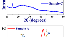
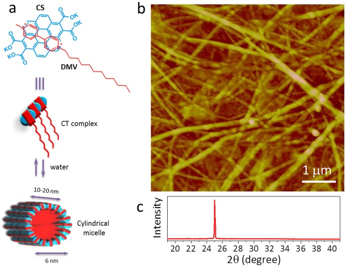
The charge transfer (CT) nanofibres were prepared by the self-assembly of coronene-viologen based donor and acceptor (D and A) pairs in water 43 (see Methods). Briefly, the potassium salt of coronene tetracarboxylate (CS) and the dodecyl substituted unsymmetric viologen derivative (DMV) are used as D and A pairs which interact via ground-state CT interactions to form a hierarchical self-assembly. In water, these two components stack themselves to form cylindrical micelles (diameter, ≈6 nm) following a surfactant-like assembly (schematic in Fig. 1a). This forms bilayers of CT-amphiphiles arranged radially with the D and A molecules stacked face-to-face along the length of the nanofibre. The tapping mode AFM topography image in Fig. 1b shows nanofibres crystallised from 1 mM solution, of varying diameters (100–300 nm) interwoven randomly to form a carpet morphology. The XRD pattern of the carpet film in Fig. 1c shows a peak at 2θ ≈ 25.031° corresponding to a d-spacing of 3.5545 Å. Given the constituents in the nanofibre, this value should correspond to the π – π spacings in the D and A pairs along the nanofibre. The intensity and sharpness associated with the peak indicate highly ordered nature of the D and A stacking.
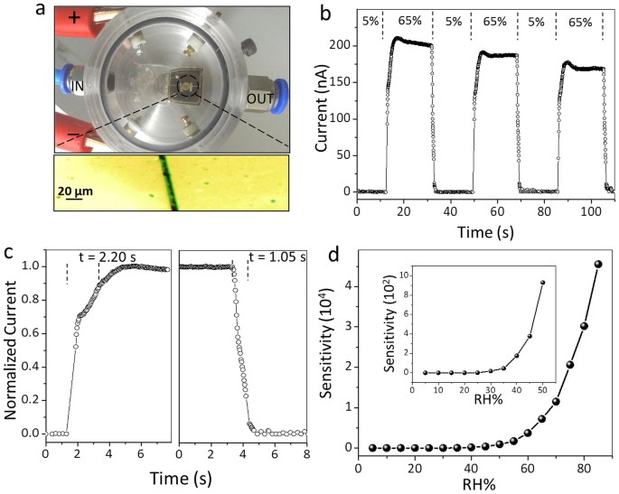
The performance of the nanofibres as a humidity sensor is described in Fig. 2. A drop (5 μL, 1 mM) of the CS-DMV nanofibre solution was drop coated on gold gap electrodes on glass and allowed to dry for an hour. The device was then placed inside a humidity cell (Fig. 2a) and connected to a Keithley 4200 source unit for electrical characterisation. Moisture-nitrogen mixture of varying RH was passed through the humidity cell through the inlet as shown in Fig. 2a. As the RH increased from 5% to 65% at 0.8 V bias, the current in the nanofibres increased from ≈0.1 nA to 220 nA (Fig. 2b) and showed a small decay while RH was still on, but dropped down to 0.1 nA sharply when RH switched to 5%. The response and recovery times, measured as the time taken to reach 90% of the peak value (Fig. 2c), are 2.20 and 1.05 s respectively. The sensitivity of the device, which is a direct measure of how well a device detects per unit change of RH, is defined in this case as the change in current normalized with respect to the current at 5% RH 9 . The sensitivity increased rapidly as RH increased to 85% to reach a value of ≈45,000 (Fig. 2d, for current values see Supplementary Fig. S1). The fabricated devices were stable over several months when stored under ambient conditions (see Supplementary Fig. S2). The effect of environment temperature (between 10°C to 50°C) on the sensor has also been studied and described in the Supplementary Fig. S3.
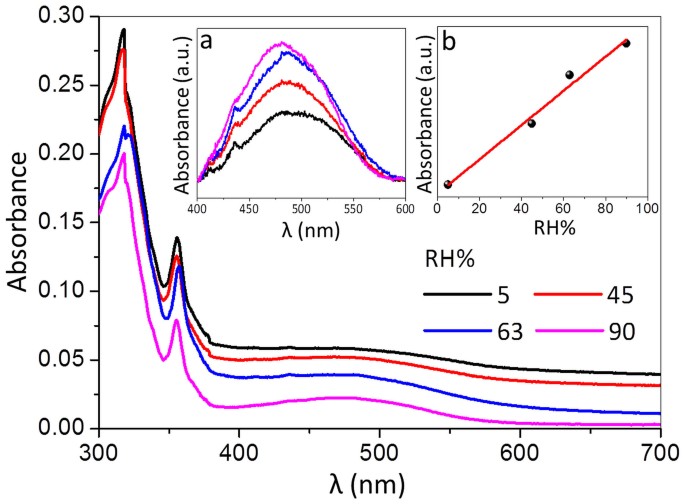
In order to gain an insight into the observed electrical changes with RH, UV-vis absorption measurements were carried out on a CS-DMV nanofibre carpet film as shown in Fig. 3. Few drops of the nanofibre solution were drop coated on the inner side of a quartz cuvette and allowed to evaporate. The cuvette was then closed with a rubber stopper hosting inlet and outlet for passing moisture-nitrogen mixture of varying RH. The absorption spectra of CS-DMV nanofibres film exhibit strong absorption peaks at 318 and 355 nm along with a broad band centred around 480 nm which originates from the ground state intermolecular charge transfer interaction between donor and acceptor molecules 43 . The peak intensity is highly sensitive to any perturbation to the charge transfer interaction, which in turn influences the electrical transport through the nanofibre. The intensity of the 480 nm band does show changes with change in RH (Fig. 3), but accompanying this change, the spectral baseline also shifts due to varied transmittance of the film (vide infra). After baseline correction, the changes in the intensity of the 480 nm band became quite apparent (see inset (a) of Fig. 3). Indeed as shown in inset (b), the intensity was found to increase linearly with increase in RH emphasizing the role of charge transfer in electrical transport of CS-DMV nanofibre system.
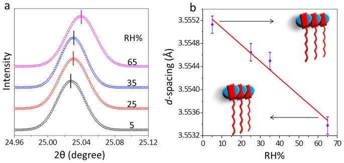
Further insight into the structural changes comes from in situ X-ray diffraction measurements (Fig. 4). With increase in RH from 5% to 65%, the peak corresponding to the CS and DMV spacing (π-π interaction distance) in the supramolecular assembly is found to shift from a 2θ value of 25.027° (d-spacing, 3.5551 Å) to 25.040° (3.5534 Å), the peak shift of 0.016 Å being well outside of the measurable limit (±0.00015 Å). The peak shift is linear (Fig. 4b) and thus follows a similar behaviour as the intensity of the charge transfer peak (Fig. 3b). Thus it appears that an increase in RH brings the CS and DMV units closer, thereby enforcing a much tighter packing in the assembly. Since the electrical transport in such systems depend sensitively on the charge transfer interaction and π- π delocalization among the D and A pairs 44 , any small perturbation due to change in RH can bring about considerable change in conductivity.
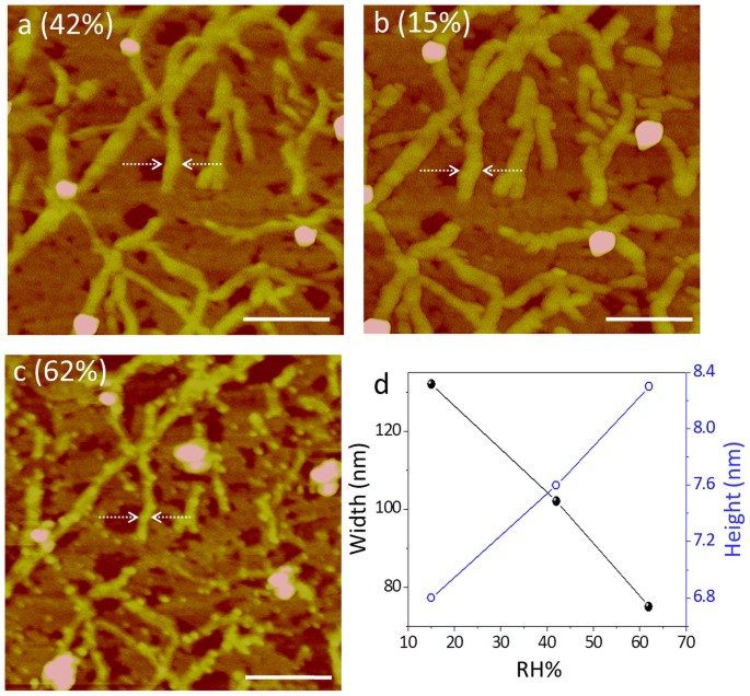
The local changes in the nanofibre assembly as discussed above, may have some implications on the morphology of the nanofibres. AFM measurements were carried out on the nanofibre film at different RH values in an environmental hood (see Fig. 5 and Supplementary Fig. S4). At ambient humidity (42% RH), the average width of the nanofibres was 99.6 ± 7.6 nm (Fig. 5a) which increased to 118.4 ± 9.6 nm when the RH in the hood was changed to 15% (Fig. 5b). The concomitant change in nanofibre height was from 6.24 ± 1.1 to 5.2 ± 1.18 nm. As the humidity was increased to 62%, the average width decreased to 76.4 ± 9.0 nm while the height increased to 8.14 ± 0.5 nm (Fig. 5c). The decreasing width can enhance light transmission as seen from the downward shift of the spectral baseline in Fig. 3a. In Fig. 5d are shown the observed morphology changes with respect to a given location on a nanofibre (see arrow marks in Fig. 5a, b and c). Many such individual measurements have been performed (Supplementary Fig. S2). The observed morphology changes may be taken to indicate a tighter molecular assembly with higher humidity around the supramolecular fibres. Under very dry conditions, the nanofibres were found to be non-conducting. Indeed, the conduction could be initiated by simply exposing to humid air.
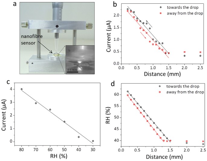
As it is clear from the above observations, the CS-DMV nanofibres can be potentially used for monitoring small variations in humidity levels near a surface, for example; such ideas could be relevant in the context of next generation intelligent device components such as artificial skin or touchless screen, where the ‘sensation’ of an approaching finger or a live cell would depend on its distance from the skin 15 . We designed an experiment wherein the change in current through a nanofibre film was monitored while a microtip carrying a tiny water droplet made a controlled approach (see Fig. 6a), from a distance of 2.5 to 0.2 mm in steps of 0.1 mm. As seen from the plot in Fig. 6b, the current in the circuit increased from ≈0.45 to 2.3 μA and the increase was nearly linear with distance. On retracting the tip, it followed a similar trend with slightly less current. Based on the calibration data collected for this film (Fig. 6c), we were able to estimate RH at different tip separation distances (Fig. 6d). It is interesting to see that the RH value reaches as high as 60% close to the drop and decreases gradually till 1.5 mm above which it merges with the ambient background. This trend is similar to the one reported by Feng et.al. 15 , although this study aims at measurements much closer to the humid surface. This experiment clearly demonstrates the ‘action at a distance’ possibility in real applications with high degree of sensitivity.
It may be noted that the nanofibre carpet device developed in this study exhibits quite fast response (≈1–2 s) comparing well with many literature examples (see Supplementary Table S1). It is known from the literature that conducting paths in carpet-like morphology come across innumerable scattering junctions, which can adversely influence the device response 25,26,27 . We have explored improving the response by fabricating a device with only few nanofibres across the gap electrodes instead of a carpet film. This was achieved by placing a 5 μL drop of 0.1 mM CS-DMV dispersion (Fig. 7a). The change in the voltage across a standard resistor (20 MΩ) as measured using an oscilloscope was quite rapid (Fig. 7b) as the humidity over the device was switched between 5% and 80% in cycles. In this case, the response and recovery times turned out to be 8 and 24 ms respectively (Fig. 7c). To our knowledge, this is the fastest humidity sensor made so far; nafion persulphonate 4 and graphene oxide 17 based sensors come close to its performance with 30–40 ms response time (see Supplementary Table S1). The sensitivity of the device was in the range, 100–500 and its variation at different RH is shown in Supplementary Fig. S5.
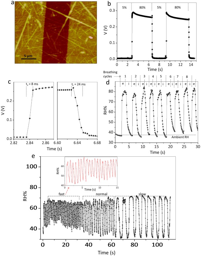
Motivated by the fast response and sensitivity of the sensor, we found it interesting to explore the usefulness of this device in monitoring human breath, particularly during exhale as the humidity is expected to be different (usually higher) compared to inhaled (ambient) air. Working with a healthy adult male, the variations in the RH value were monitored by holding the nanofibre device at a distance of 3.5 cm from the nose while breathing normally. Interestingly, the RH over the device showed sharp rise during exhaling and dropped to ambient value while inhaling, which was nearly repetitive corresponding to the breathing cycles (see Figure 7d). Further, the device could efficiently follow, no matter how fast or slow was the breathing (Figure 7e). In the panel given, the breathing varied from 0.3 to 5 s per cycle. Importantly, the nanofibres in the device were stable throughout.
Such a device should be of importance in breath analysis. Characterization of exhaled breath has become a simple yet powerful non-invasive method of diagnosing diseases such as asthma, cancer, diabetes etc 45,46,47,48,49,50 . Any serious monitoring of exhaled breath should be an online process tracking the changes in every breath cycles 51,52 ; off-line trace gas analysis suffers from inherent problems associated with sample preparation and storage, contamination, chemical transformation of the analytes, etc 52 . While monitoring analytes for specific diseases is important, monitoring humidity itself can possibly throw some light on an individual's health such as dehydration. Although there are many indices suggested in the literature to indicate body hydration status 53,54,55 , we for the first time, have made an attempt monitor dehydration through RH in the breath.
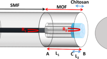
Towards this end, we considered it important to measure simultaneously the humidity and the exhaled breath flow, the latter to increase the confidence in the measurements. We therefore, fabricated a flow cum humidity sensor using CS-DMV nanofibres (Fig. 8). The device consists of supramolecular humidity sensors, D1 and D2, placed in a glass tube 8 cm apart. The glass tube is connected to a breath receiving mask (typically used for oxygen supply to patients), through a flow divider (see Fig. 8a); the latter was required as calibration using typical mass flow controllers proved difficult at realistic breath flow (≈10–50 ksccm 56 ). As an individual breaths out into the mask, the divided flow reaches the sensor D1 at time t1 and D2 at time t2 (at half rise) and the time difference, Δt = t2 − t1 (see Fig. 8b) was used for calculating the flow. The humidity could be measured from both sensors using the respective calibration curves (see Supplementary Fig. S6). By measuring both humidity and flow rate for every breath cycle, the humidity measurement could be validated.
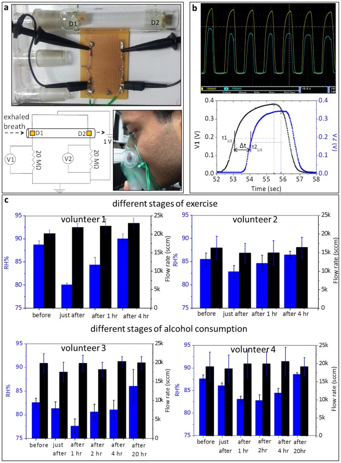
The above setup was used in varied conditions of volunteers. We have monitored the RH present in nasal breath rather than that from mouth 34 , as the former can be considered as a better representative of the lung hydration levels (with no interference from mouth saliva). The volunteer was made to breath comfortably but consistently such that the flow rate of breath was in a reasonably narrow range (20–30 ksccm). The data shown in Fig. 8c were collected from four volunteers. Average RH and flow rate values obtained for volunteer 1 (male) in normal conditions were 88.8 ± 0.8% and 20.2 ± 0.8 ksccm respectively (see histogram in Fig. 8c and Supplementary Fig. S7a). The volunteer was then made to run around 10 km in nearly 1 hr. After relaxing for few minutes, both the quantities were measured again (see histogram in Fig. 8c). It is interesting to see a sharp decline in the RH value in the exhaled breath after exercise (80.1 ± 0.3%), which is understandable as the volunteer got dehydrated due to perspiration. The volunteer was indeed feeling thirsty, but was restrained from consuming food and water. When measurements were done after 1 hr, surprisingly, we found a gradual recovery of RH in the breath (84.4 ± 1.6%). It appears that water from the internal body fluid was diverted towards the respiratory system which reflects in the breath RH level returning to normalcy (90.1 ± 1.1%, see histogram in Fig. 8c). Human lung tissue is known to maintain high degree of wetness for efficient exchange of gases and when dehydration occurs, it is susceptible to dry out thereby reducing the water content in the exhaled breath. However in our study, we have found that RH in the breath regained even under the dehydrated condition, clearly indicating that the lung tissue, by its nature, regenerated moisture on its (alveolar) surface from various metabolic processes. Upon water intake after the 1 hr measurement, the RH value completely regained (Fig. 8c). As shown on the right in Fig. 8c, a similar but mild trend was observed in the case of a female volunteer who undertook to run 3 km in nearly 30 minutes (see Fig. 8c and Supplementary Fig. S7b).
Another important situation of body dehydration relates to alcohol consumption 57 . Here, we examined the relative humidity in the exhaled breath to see the effect of alcohol consumption (but not as alcohol sensor). In the given two instances (Fig. 8c, bottom), the male volunteers 3 and 4 after consuming alcohol (beer, 8% alcohol, 2 L) were restrained from consuming food and water. We observed a decline in the RH following alcohol consumption due to dehydration in the body. However, the RH values recovered in spite of not feeding water (see bottom histograms in Fig. 8c and Supplementary Fig. S8). The variation in breath alcohol concentration (BAC) was monitored at every stage; RH declined with increased BAC (Supplementary Table S2). The situation is similar to that induced by physical exercise. Among many other control experiments, we have also monitored the RH of exhaled breath when a volunteer was made to drink plenty of plain water to cause excessive hydration; indeed, it was well reflected in the breath RH measurements (see Supplementary Fig. S9). Thus in this study, we have measured both flow and RH for every cycle where flow in narrow range ensures good sampling, removing other influences on RH. While this is a preliminary study of body dehydration monitored through RH in the breath, the study should open up new insights into diagnosis through breath analysis.
An ultra-fast, wide range, highly sensitive resistive humidity sensor has been developed based on a coronene-viologen derivative system, self-assembled in the form of nanofibre from water. While a film of nanofibres exhibited high sensitivity (≈10 4 ), devices with few nanofibres showed extremely fast response (≈10 ms). Using UV-vis, XRD and AFM measurements, we have found that the π-π interaction between the donor and the acceptor molecules depends sensitively on the surrounding humidity influencing electrical conduction across the nanofibre. The fabricated devices were found to be stable over 8 months during the study. As is evident, these devices can be used in various contexts. The RH value close to a drop of water was determined by approaching it with a nanofibre film sensor. Fast response devices made of few nanofibres have been effectively employed for dynamic monitoring of human breath. Using two such devices in a circuit, a flow cum humidity sensor has also been fabricated and its application has been demonstrated in analysing dehydrated conditions. As the sensing element is a simple organic system from solution processes, these devices are essentially of low cost and also environment friendly. Potential applications in personal healthcare as use-and-dispose devices, is evident.
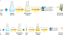
The detailed procedure can be found in Reference 43 . In brief, CS (coronene tetracarboxylate) was synthesized by a twofold oxidative benzogenic Diels–Alder reaction of perylene with N-ethyl maleimide and subsequent hydrolysis with KOH in methanol. DMV (dodecyl methyl viologen) was synthesized from 4,4′-bipyridine by controlled reaction on one nitrogen with dodecyl bromide to give mono pyridinium ion and followed by treating the with methyl iodide to amphiphllic dicationic bipyridine (DMV). The charge-transfer fibres have been assembled from injection of a methanol solution of DMV, in which the viologens are molecularly dissolved, to the aqueous solution containing free CS molecules (10% v/v methanol in water).
The structural analysis was carried out by X-ray diffraction using Bruker D8 Discover diffractometer attached with temperature and humidity controlled stage. Cu Kα (λ = 1.5419 Å) was used as X-ray source. Atomic force microscopy (AFM) was performed using Multimode, Veeco digital instruments, USA with Nanoscope IV controller. The humidity control was achieved by using an environmental hood with flow of water vapors of different RH 15%, 42% and 62% continuously for 15 minutes. The images were collected in the tapping mode using Si cantilevers. The FE-SEM imaging was done using a Nova NanoSEM 600 (FEI Co., The Netherlands) by drop casting the aqueous solution on Si(100) substrate followed by drying in vacuum and was operated with an accelerating voltage of 10 kV. Absorption spectra were recorded on a Perkin Elmer Lambda 900 UV-VIS-NIR Spectrometer using 1 mm path length cuvette. To analyse the intensity variation with increasing RH, a baseline was drawn for each spectrum using Origin 8.0 software in the region of interest, 400 to 600 nm. The raw data was then subtracted from the baseline. Vapors of different RH were obtained by controlling the equilibrium vapor pressure of sulphuric acid 15 . A humidity cell was constructed for rapid switching of RH and switching between two RH levels was done using a Tee with flow rate of 200–300 sccm. A commercial humidity meter Testo 410-2 was used to measure the obtained RH. For alcohol monitoring a commercial alcohol meter (BAC TRACK B70) was used. We confirm that the Ethical Committee of the JNCASR approved all the experiments described in this paper. Further, informed consent was obtained from all the subjects.
The glass substrates were cleaned in piranha solution followed by washing in distilled water several times. Metallic contacts were made by physical vapor deposition of Au by shadow masking using a resistive thermal evaporator (HindHivac, India) at a base pressure of 10 −6 Torr. A Keithley Semiconductor Characterization System 4200 was used to measure device characteristics. An Oscilloscope Tektronix DPO 4104 was used to measure the voltage drop across resistors.
The authors thank Prof. C.N.R. Rao for his constant encouragement. They thank Mr. K. Venkata Rao for synthesising the molecules used in the study. They are thankful to the volunteers for their participation in data collection. This research work is supported by Department of Science and Technology (DST), New Delhi, India. A.A.S. is thankful to DST for Nano-Postdoc Fellowship. U.M. thank CSIR, India for a research fellowship.